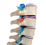
Strap in folks, it’s time for an anatomy lesson! It has been a long time since I sat in my human biology class studying the inner working of the human spinal cord and I’m guessing it’s been a while for you, too! The below article paints a really vivid picture of the parts of the spinal cord and their functions. Enjoy!
How does the spinal cord work?
“The spinal cord is a tight bundle of neural cells (neurons and glia) and nerve pathways (axons) that extend from the base of the brain to the lower back. It is the primary information highway that receives sensory information from the skin, joints, internal organs, and muscles of the trunk, arms, and legs, which is then relayed upward to the brain. It also carries messages downward from the brain to other body systems. Millions of nerve cells situated in the spinal cord itself also coordinate complex patterns of movements such as rhythmic breathing and walking. Together, the spinal cord and brain make up the central nervous system (CNS), which controls most functions of the body.”
“The spinal cord is made up of neurons, glia, and blood vessels. The neurons and their dendrites (branching projections that receive input from axons of other neurons) reside in an H-shaped or butterfly-shaped region called gray matter. The gray matter of the cord contains lower motor neurons, which branch out from the cord to muscles, internal organs, and tissue in other parts of the body and transmit information commands to start and stop muscle movement that is under voluntary control. Upper motor neurons are located in the brain and send their long processes (axons) to the spinal cord neurons. Other types of nerve cells found in dense clumps of cells that sit just outside the spinal cord (called sensory ganglia) relay information such as temperature, touch, pain, vibration, and joint position back to the brain.”
“The axons carry signals up and down the spinal cord and to the rest of the body. Thousands of axons are bundled into pairs of spinal nerves that link the spinal cord to the muscles and the rest of the body. The function of these nerves reflects their location along the spinal cord.”
“The outcome of any injury to the spinal cord depends upon the level at which the injury occurs in the neck or back and how many and which axons and cells are damaged; the more axons and cells that survive in the injured region, the greater the amount of function recovery. Loss of neurologic function occurs below the level of the injury, so the higher the spinal injury, the greater the loss of function.”
“A whitish mixture of proteins and fat-like substances called myelin covers the axons and allows electrical signals to flow quickly and freely. Myelin is much like the insulation around electrical wires. It is formed by axon-insulating cells called oligodendrocytes. Because of its whitish color, the outer section of the spinal cord—which is formed by bundles of myelinated axons—is called white matter.”
“The spinal cord, like the brain, is enclosed in three membrane layers called meninges: the dura mater (the tougher, most protective, outermost layer); the arachnoid (middle layer); and the pia mater (innermost and very delicate). The soft, gel-like spinal cord is protected by 33 rings of bone called vertebrae, which form the spinal column. Each vertebra has a circular hole, so when the rung-like bones are stacked one on top of the other there is a long hollow channel, with the spinal cord inside that channel. The vertebrae are named and numbered from top to bottom according to their location along the backbone: seven cervical vertebrae (C1-C7) are in the neck; twelve thoracic vertebrae (T1-T12) attach to the ribs; five lumbar vertebrae (L1-L5) are in the lower back; and, below them, five sacral vertebrae (S1-S5) that connect to the pelvis. The adult spinal cord is shorter than the spinal column and generally ends at the L1-L2 vertebral body level. A thick set of nerves from the lumbar and sacral cord form the “cauda equina” in the spinal canal below the cord.”
“The spinal column is not all bone. Between the vertebrae are discs of semi-rigid cartilage and narrow spaces called foramen that act as passages through which the spinal nerves travel to and from the rest of the body. These are places where the spinal cord is particularly vulnerable to direct injury.”
(Source https://www.ninds.nih.gov/)
If you or a loved one is dealing with any of the complications related to a spinal cord injury at work or if you would like more information on the Virginia workers’ compensation system, order my book, “The Ultimate Guide to Workers’ Compensation in Virginia,” or call our office today (804) 755-7755.
~Author
Michele Lewane, Esq.
About the Author: Michele Lewane
The Injured Workers Law Firm is a Richmond, Virginia based firm solely focused on serving clients with workers' compensation claims in Virginia. If you have questions about your benefits or if you would like more information on the Virginia workers’ compensation system, order our book, “The Ultimate Guide to Workers’ Compensation in Virginia” , or call our office today (804) 755-7755.


